 |
The Inside Story
II. Compositional Tomography |
|
|
The Inside Story
II. Compositional Tomography |
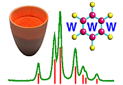
Compositional Tomography of Mineral and Engineering Objects
10 to 15 years ago the prospect of examining large dense objects was considered
to be feasible only with neutrons; synchrotron sources have changed all that.
In the Diffraction II section we noted that whereas some common materials could
only be penetrated by distances of around 50 µm in the case of
laboratory X-rays (e.g. copper Kα 1.54 Å), such penetration
increases to the millimetre level at shorter wavelengths (e.g.
0.56 Å). However third generation synchrotron multipole magnet
devices produce intense X-rays with energies more than an order of magnitude
greater than the laboratory copper source. This has enabled many experiments to
be performed on the internal analysis of bulk objects such as rock minerals,
building stone and concrete, ceramic/metal objects and so on. There is space to
describe just one technique with one example: that is, looking at the interior
of a large
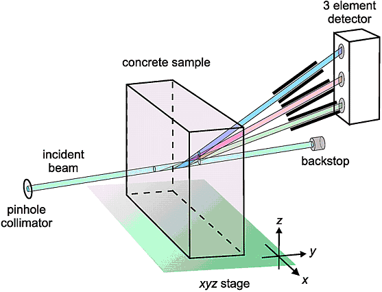
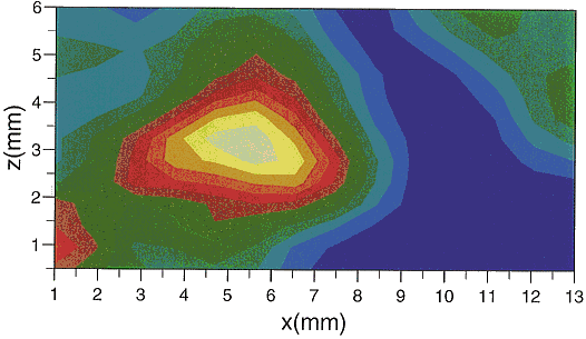
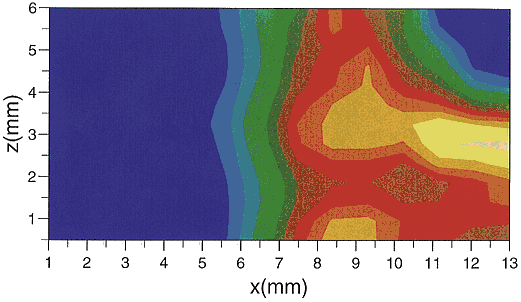
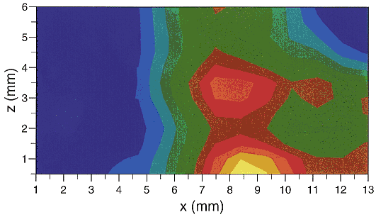
A key feature again is that the region of interest inside the concrete is defined by the collimated incident and diffracted beams. The concrete block is traversed in a raster fashion (in this particular case in two dimensions; i.e. along the x- and z-directions) so that the diffracting region covers a region of interest; in this case a 13 × 6 mm area covered in 13 × 13 traverse steps. The beauty of energy-dispersive diffraction is that the whole diffraction pattern is collected by a stationary energy-dispersive detector. Another twist in this case is that 3 detectors were used (see previous diagram) so 3 diffraction patterns, which complemented each other, were collected each time from the same region. Eventually diffraction patterns were collected from all points over the traversed volume and so one could then construct maps of various phases according to the intensity of their characteristic diffraction peaks. In the following figure we show 4 such maps for the traversed 13 × 6 mm area.
| Calcite | Dolomite |
 |
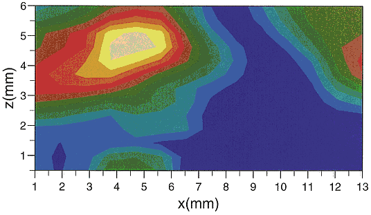 |
| Portlandite | Ettringite |
 |
 |
|
© Copyright 1997-2006.
Birkbeck College, University of London.
|
Author(s):
Paul Barnes Simon Jacques Martin Vickers |