 |
Mode (1): Flat Plate with/without Analyser Crystal |
|
|
Mode (1): Flat Plate with/without Analyser Crystal |
Mode (1): Flat Plate with/without Analyser Crystal
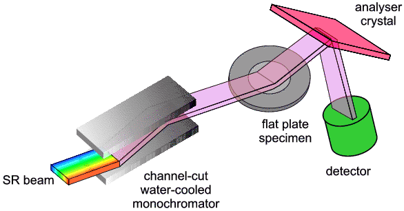
The diagram below illustrates this basic mode (without analyser crystal): a beam of near-parallel monochromatic X-rays strikes the surface of a flat "plate" sample at an angle θ. If the Bragg condition is satisfied and diffraction occurs, as illustrated, then the diffracted X-rays which reach the detector will also be travelling at an angle θ to the sample; this means that the total angle between the incident and diffracted beams is 2θ. This configuration can be retained for different θ provided we always rotate the detector twice as much (2θ) as the sample (θ). There are some advantages in using this so-called 2θ:θ motion, and you will come across it often during this course.
It is important to understand that higher density materials, including many inorganic compounds, are highly absorbent of x-rays at the wavelengths in common use for powder diffraction. As a result of this, where flat plate sample geometry is in use most of the diffraction occurs within a thin layer close to the sample surface. One consequence of this is that, on average X-ray photons travel a similar distance within the sample before and after they are diffracted (this will be explained more fully later in the course). It turns out that this gives rise to an advantageous "cancelling out" feature, making the effects of X-ray absorption much less of a problem. A disadvantage, however, of flat-plate holders is that they can give rise to preferred orientation.
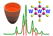
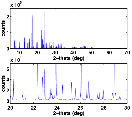
The diagram is shown together with a typically good quality powder diffraction pattern from fluorescein diacetate, collected over 6 hours on Daresbury SRS-synchrotron station 9.1.

|
© Copyright 1997-2006.
Birkbeck College, University of London.
|
Author(s):
Paul Barnes Simon Jacques Martin Vickers |