 |
Powder Diffraction Project
byStewart F. ParkerISIS Facility, Rutherford Appleton Laboratory Chilton, Didcot, Oxon OX11 0QX, UK |
 |
|
Powder Diffraction Project
byStewart F. ParkerISIS Facility, Rutherford Appleton Laboratory Chilton, Didcot, Oxon OX11 0QX, UK |
|
Introduction
The aim of the project was to provide some experience of crystal structure refinement from powder data by the Rietveld method. Angle-dispersive powder neutron diffraction data was chosen since there are fewer problems due to texture or preferred orientation than would probably be encountered with X-ray data.
Experimental
A powder neutron pattern of lead sulfate was collected at 5 K on the high-resolution powder diffractometer, D2B, at the Institut Laue Langevin, (Grenoble, France). A nominal wavelength of 1.594 Å was used and the data collected between 8 £ 2q £ . 142.975° with a step length of 0.025°. Data files A and B in the 2q, intensity, and s format were obtained from http://pd.cryst.bbk.ac.uk/pd99/assign. For the analysis, the program LHPM-Rietica was downloaded from the CCP14 mirror site.
Results
Part 1: Structure refinement using PbnmAt room-temperature the structure of lead sulfate is orthorhombic, space group P bnm (number 62). As initial values the refined parameters from the case study were used:
Cell parameters : a = 6.94802 Å, b = 8.46618 Å, c = 5.38904 Å
Atom List |
x |
y |
z |
Pb |
0.1671 |
0.1879 |
1/4 |
S |
0.1848 |
0.4358 |
3/4 |
O(1) |
0.0958 |
0.5907 |
3/4 |
O(2) |
0.0429 |
0.3068 |
3/4 |
O(3) |
0.3082 |
0.4192 |
0.9727 |
We are told that the peakshape is approximately Gaussian so this was used as the starting point and initial values of UVW used were 0.05, -0.15, 0.20 (°2) as suggested.
The refinement followed the strategy employed in the case study. A polynomial background was used, it was found that additional terms after 3 gave no significant improvement in the fit. Sequential addition and refinement of: the scale factor, cell parameters, zero error, (x,y,z) of O(3), (x,y) of Pb, (x,y) of O(1) and O(2), (x,y) of S, overall B factor and finally of UVW and the Lorentzian contribution to the lineshape were carried out. The final values are given in Table 1 and a plot of the final fit is shown in Figure 1.
R-Factors : Rp = 4.05, Rwp = 5.21, Rexp = 2.97 and c 2 = 3.08 Cell parameters : a = 6.9261(4) Å, b = 8.4152(5) Å, c = 5.3684(3) Å
B overall : 0.32 Å2 Instrumental : U = 0.7185, V = -0.5729, W = 0.2926 (°2), % Lorentz = 10.8, 2q error = 0.069 ° Structural Parameters :
(The " ¢ " indicates that the coordinates of this atom are symmetry related). |
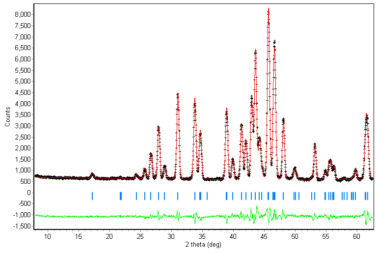
Figure 1: Comparison of data (black crosses), and the fit (red line) for the small data set assuming space group Pbnm . The calculated hkl positions (blue bars) and the difference spectrum (green line) are also shown.
From the quality of the fit shown in Figure 1 and the low R-factors obtained, this would appear to be an excellent result. The parameters obtained are reasonable; in particular, the cell dimensions have decreased from the room temperature values as would be expected on cooling. The only surprising note is that the sulfate bond lengths and angles are significantly different from those at room temperature. This is unexpected, since the geometry would not be expected to change dramatically with temperature because the intramolecular forces are much stronger than the intermolecular ones. This is a hint that perhaps something is not right. This is confirmed in Figure 2 which was obtained using the full data set with the same parameters.
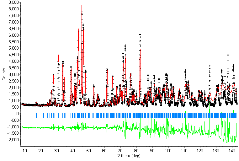
Figure 2: Comparison of data (black crosses), and the fit (red line) for the full data set assuming space group Pbnm . The calculated hkl positions (blue bars) and the difference spectrum (green line) are also shown.
R-Factors : Rp = 17.74, Rwp = 29.49 and c 2 = 106.32
The R-factors and the fit displayed in Figure 2 show that the model is actually very poor, despite the excellent fit to the small data set. This clearly demonstrates the need for high angle data. The reason this is essential is also shown in Figure 2; there are very many more reflections per degree at high angles than at low angles so most of the information in the diffraction pattern is at high angles.
Part 2: Structure refinement using P21/n
The room-temperature structure of lead sulfate has the space-group symmetry Pbnm in which the sulfate anion has mirror-plane symmetry. One possible reason for the result observed in the preceding refinement is that a slight distortion of the unit cell occurs with consequent loss of the mirror-plane symmetry on cooling the sample to 5 K. The two lower-symmetry space groups of Pbnm that will need to be considered are therefore (i) P21/n and (ii) P21/b (with the symmetry elements relating to the x-axis). These are actually the same space group, but with different arrangements of the symmetry elements with respect to the unit-cell axes.
In both of these space groups, the atoms that formerly occupied special positions (Pb, S, O(1) and O(2)) in Pbnm, now occupy general positions, thus x,y,z are all refinable for them. In addition, the loss of the mirror plane means that the symmetry equivalent position of O(3) (called O(4) from now on) now needs to be specified. As starting parameters, I have used the refined positions and instrumental parameters from the Pbnm refinement above with the additional atomic position for O(4):
O(4) atomic coordinates : x = 0.3070(1), y = 0.4184(1), z = 0.5250
The refinement followed the strategy used previously. A three-term polynomial background followed by sequential addition and refinement of: the scale factor, a, b and c cell parameters, zero error, (x,y,z) of O(3) and O(4), (z) of Pb, S, O(1) and O(2), (x,y) of Pb, S, O(1) and O(2), b, overall B factor, UVW and the Lorentzian contribution to the lineshape and isotropic B factors were carried out. The final values are given in Table 2, a plot of the final fit is shown in Figure 3 and the unit cell in Figure 4. A powder CIF file is attached.
| Table 2:
Final parameters for the refinement of the large data set in space group P 21/n R-Factors : Rp = 4.34, Rwp = 5.59, Rexp = 2.85 and c 2 = 3.84 Cell parameters : a = 6.9282(1) Å, b = 8.4189(1) Å, c = 5.3714(1) Å, b = 89.7619(7) °
Instrumental : U = 0.04658, V = -0.14029, W = 0.20719 (°2), % Lorentz = 16.33, 2q error = 0.092 ° Structural Parameters :
|
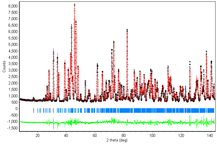
Figure 3: Comparison of data (black crosses), and the fit (red line) for the full data set assuming space group P21/n . The calculated hkl positions (blue bars) and the difference spectrum (green line) are also shown.
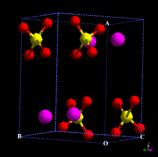
Figure 4: Unit cell of PbSO4 at 5 K assuming spacegroup P21/n .
Part 3: Structure refinement using P21/a
I was unable to refine the structure in P21/b, so I adopted the suggestion to refine in P21/a by interchange of the x and y coordinates. The starting parameters were therefore:
Cell parameters : a = 8.4152 Å, b = 6.9261 Å, c = 5.3712(1) Å
Atom List |
x |
y |
z |
Pb |
0.1891(2) |
0.1678 |
1/4 |
S |
0.4317(8) |
0.1933 |
3/4 |
O(1) |
0.5921(4) |
0.0926 |
3/4 |
O(2) |
0.3075(4) |
0.0394 |
3/4 |
O(3) |
0.4184(2) |
0.3070 |
0.9753 |
O(4) |
0.4184(2) |
0.3070 |
0.5250 |
The refinement followed exactly the same pattern as for refinement in P21/n. The final values are given in Table 3, a plot of the final fit is shown in Figure 5 and the unit cell in Figure 6. A powder CIF file is attached.
Table 3: Final parameters for the refinement of the large data set in space group P 21/a R-Factors : Rp = 9.95, Rwp = 13.72, Rexp = 2.85 and c 2 = 23.18 Cell parameters : a = 8.4187(2) Å, b = 6.9276(2) Å, c = 5.3697(2) Å , b = 90.0645(32) °
Instrumental : U = -0.010, V = 0.054, W = 0.155 (°2), % Lorentz = 19.38, 2q error = 0.084 ° Structural Parameters :
|
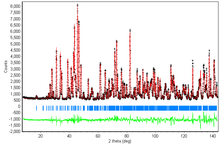
Figure 5: Comparison of data (black crosses), and the fit (red line) for the full data set assuming space group P21/a . The calculated hkl positions (blue bars) and the difference spectrum (green line) are also shown.
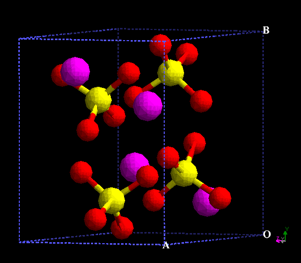
Figure 6: Unit cell of PbSO4 at 5 K assuming spacegroup P21/a .
Conclusions
Of the two possible low temperature models the P21/n structure is superior in terms of the R-factors and the estimated standard deviations (esd) of the parameters. The latter are twice as large for the fractional coordinates in P21/a as in P21/n giving bondlengths with esd's twice as large and bond angles with esd's between two and four times as large. The pattern of S-O bondlengths and O-S-O angles is also different in both refinements with a greater similarity to the room temperature values for P21/n. Thus the bondlengths show three S-O distances approximately the same and one different in both cases, although the 'odd one' is shorter at room temperature rather than longer as seen at low temperature. This may be a result of the decreased thermal motion at low temperature. For the O-S-O angles, the pattern is one large angle, ~ 113 °, and five angles at ~108 - 109 °, exactly as seen with the P21/n refinement but not the P21/a one. In both cases, the cell parameters decrease as would be expected with the reduction in temperature. The most noticeable difference between the two refinements are the values of UVW, for P21/n they are close to the initial values, whereas for P21/a they are very different. It is possible that they are being used as 'sink' for errors in the refinement caused by the poor choice of model.
Both refinements were very sensitive to the value of b, it was only possible to refine it late in the refinement process, or unstable refinements resulted. In neither case was it possible to refine anisotropic thermal displacement parameters because unstable refinements resulted every time. This was despite attempts to refine the atoms one at a time (with isotropic parameters for the remaining atoms), in various combinations or all at once.
Out of curiosity, I also refined the room temperature structure with the complete data set. The resulting R-factors were very similar to those obtained for the P21/a refinement. Thus all the evidence suggests that the P21/n model is the better one.