Project - Sian Davies
EXPERIMENTAL -DATA SET A
Data for the sample of PbSO4were collected at 5 K on the high-resolution powder diffractometer D2B, at the ILL, Grenoble, using neutrons of wavelength 1.594 Å. Scattering lengths for Pb, S and O were 0.940170, 0.284700 and 0.580300 respectively. The instrumental resolution function parameters were initially set to U = 0.05, V = -0.15 and W = 0.20°2. The monochromator correction was 1.00 and the initial extinction parameter was set to 0.00.
Refinement of the structure was performed using the Rietica Rietveld refinement program.
For data set A (8.00 to 62.475°), the structure was refined in the orthorhombic room temperature space group Pbnm (equivalent to no. 62, Pnma) with initial cell parameters set to a = 7.0, b = 8.5 and c = 5.4 Å and V = 321.3Å3. The Pb2+ cation and the S and three O atoms from the SO42- anion lie on mirror planes reducing the number of ion pairs per unit cell to 4.
Table 1.
| Atom | x | y | z | Occupancy |
|---|---|---|---|---|
| Pb | 0.15 | 0.20 | 1/4 | 0.5 |
| S | 0.20 | 0.50 | 3/4 | 0.5 |
| O(1) | 0.10 | 0.60 | 3/4 | 0.5 |
| O(2) | 0.00 | 0.30 | 3/4 | 0.5 |
| O(3) | 0.30 | 0.40 | 0.9 | 1.0 |
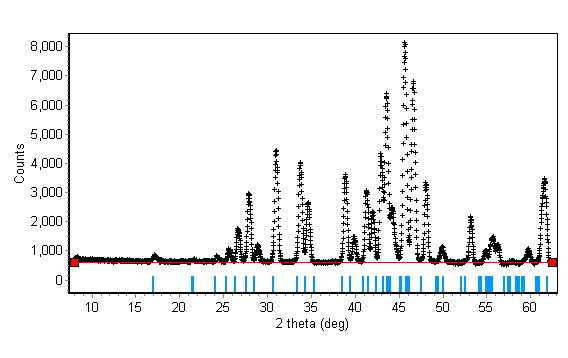
The background was estimated as 600 across the whole pattern (Fig. 1).
| Fig. 1 |
|---|
The Pseudo-Voight profile function was chosen, using the Howard, sum of 5 peaks, asymmetry, where the pseudo-Voight peak shape was 0.20 + 0.0*2q + 0.0*2q2. The data were not corrected for absorption.
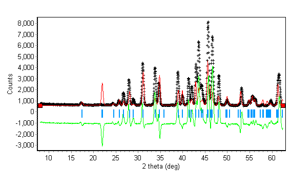
One cycle of refinement was performed to refine the overall scale factor (initially set to 0.01); the value refined to 0.48(4) (Fig. 2).
This gave figures of merit Rp = 49.92, Rwp=59.59 and Rexp = 2.92, where
the profile Rp = S|yio-yic| / Syio
the weighted profile Rwp = [Swi(yio-yic)2 / Swiyio2]1/2
the expected Rexp = [N-P / Swiyio2]1/2
The 2q zero error was then refined, from 0.00 to 0.451(6), giving R values of Rp = 34.17, Rwp = 45.16 and Rexp = 2.91. The resulting profile is shown in Fig. 2.
| Fig. 2 |
|---|
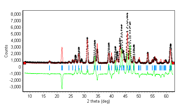
The unit cell parameters were then refined to try to improve the peak fitting. They refined to a = 6.919(3), b = 8.412(4) and c = 5.365(3)Å, with R values of Rp = 29.76, Rwp = 41.21 and Rexp = 2.91. This gave closer agreement between calculated and observed peak positions (Fig. 3).
| Fig. 3 |
|---|
The fractional coordinates of O(3) were then refined to try to improve the fit of calculated and observed intensities. They refined to a = 0.300(1), b = 0.4211(9) and c = 0.9698(12)Å with R values of Rp = 15.09, Rwp = 20.50 and Rexp = 2.91.
All general coordinates were then refined with results as in Table 2.
Table 2.
| Atom | x | y | z |
|---|---|---|---|
| Pb | 0.1672(4) | 0.1891(2) | 1/4 |
| S | 0.1982(10) | 0.4333(11) | 3/4 |
| O(1) | 0.0922(5) | 0.5918(5) | 3/4 |
| O(2) | 0.0402(8) | 0.3081(5) | 3/4 |
| O(3) | 0.3053(4) | 0.4184(3) | 0.9754(4) |
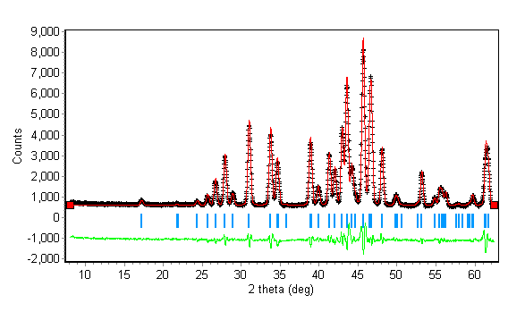
R values of Rp = 5.44, Rwp = 7.05 and Rexp = 2.90 were obtained. The profile is shown in Fig. 4.
| Fig. 4 |
|---|
Isotropic thermal parameters were then refined to try to improve the model. Originally set to 0.00, the refined values are listed in Table 3.
Table 3.
| Pb | B = 0.51(9) |
| S | B = 1.8(2) |
| O(1) | B = 0.76(13) |
| O(2) | B = 0.6(2) |
| O(3) | B = 0.64(10) |
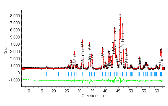
R values of Rp = 5.40, Rwp = 6.94 and Rexp = 2.90 were obtained.
The resulting profile is shown in Fig. 5.
| Fig. 5 |
|---|
The results of the refinement to this point may be summarised as follows :
Orthorhombic unit cell with a = 6.9281(5), b = 8.4165(6) and c = 5.3705(4).
Table 4.
| Atom coordinates | |||
|---|---|---|---|
| Atom | x | y | z |
| Pb | 0.1670(4) | 0.1893(2) | 1/4 |
| S | 0.1969(11) | 0.4341(14) | 3/4 |
| O(1) | 0.0922(5) | 0.5916(6) | 3/4 |
| O(2) | 0.0407(9) | 0.3082(5) | 3/4 |
| O(3) | 0.3050(4) | 0.4187(3) | 0.9748(4) |
Table 5.
| Bond lengths in Å | |
|---|---|
| S - O(1) | 1.511(10) |
| S - O(2) | 1.514(8) |
| S - O(3) | 1.427(5) |
| Bond angles in degrees | |
| O(1)-S-O(2) | 105.7(6) |
| O(1)-S-O(3) | 109.4(5) |
| O(2)-S-O(3) | 108.2(5 |
| O(3)-S-O(3') | 115.6(6) |
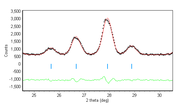
There was only a slight mismatch of the calculated and observed peak shape as shown in Fig. 6.
| Fig. 6 |
|---|
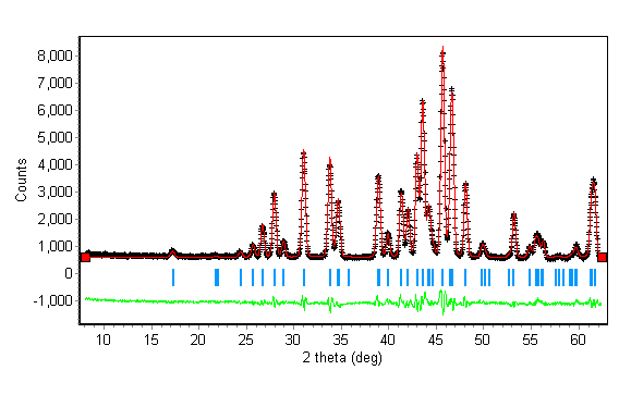
The instrument resolution function parameters were then allowed to refine, followed by refinement of the extinction parameter. They refined to U = -0.08(11), V = 0.02(10) and W = 0.15(2) and the extinction parameter refined to -0.0012(3). The peak asymmetry parameter was refined to 0.127(13) with the anisotropic Gaussian peak broadening parameter refined to 0.31(2). The resulting R values were Rp = 4.33, Rwp = 5.38 and Rexp = 2.89 (Fig. 7).
| Fig. 7 |
|---|
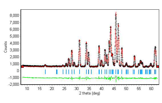
The anisotropic thermal parameters of Pb, then S, O(1), O(2) and O(3) were refined with restrictions on U13 and U23 for atoms on the mirror planes (U = 0) until convergence. The resulting R values were Rp = 4.07, Rwp = 5.03 and Rexp = 2.87 where N-P = 1871. c2 = 3.1, where c2=(Rwp/Rexp)2
The final profile is shown in Fig. 8 and the results may be summarised as follows
The overall scale factor refined to 2.80(4)
the overall temperature factor refined to -5(7)
The 2q zero error refined to 0.179(13)
The instrumental resolution function parameters refined to U = -0.00(11), V = -0.05(10), W = 0.16(2)
The peak asymmetry parameter refined to 0.124(13)
The anisotropic peak broadening parameter refined to 0.38(2)
The extinction parameter refined to -0.0031(7)
The unit cell refined to a = 6.9362(13), b = 8.4277(15), c = 5.3757(10) and V = 314.49(10)
| Fig. 8 |
|---|
The atomic parameters are listed in Table 6.
Table 6.
| Atom | x | y | z | B11 | B22 | B33 | B12 | B13 | B23 | Occupancy |
|---|---|---|---|---|---|---|---|---|---|---|
| Pb | 0.1644(11) | 0.1894(3) | 1/4 | 0.05(13) | 0.04(9) | 0.1(2) | 0.0(4) | 0.0 | 0.0 | 0.5 |
| S | 0.187(3) | 0.435(3) | 3/4 | 0.0(4) | -0.0(3) | -0.1(6) | 0.008(4) | 0.0 | 0.0 | 0.5 |
| O(1) | 0.0903(10) | 0.5913(2) | 3/4 | 0.0(6) | 0.0(4) | 0.1(10) | 0.006(2) | 0.0 | 0.0 | 0.5 |
| O(2) | 0.0444(14) | 0.3055(11) | 3/4 | 0.0(6) | 0.0(4) | 0.0(9) | -0.001(2) | 0.0 | 0.0 | 0.5 |
| O(3) | 0.3051(7) | 0.4176(4) | 0.9743(8) | 0.0(3) | 0.0(2) | 0.0(4) | -0.011(3) | -0.002(2) | -0.001(2) | 1.0 |
Table 7. shows the final molecular dimensions
Table 7.
| Bond lengths in Å | |
|---|---|
| S - O(1) | 1.47(2) |
| S - O(2) | 1.47(2) |
| S - O(3) | 1.467(12) |
| Bond angles in degrees | |
| O(1)-S-O(2) | 111.0(15) |
| O(1)-S-O(3) | 110.1(11) |
| O(2)-S-O(3) | 107.5(11) |
| O(3)-S-O(3') | 110.6(12) |
EXPERIMENTAL -DATA SET B ; space group P21/n
On cooling the sample to 5K, there is a distortion in the unit cell and a consequent change in the crystal system with the loss of the mirror plane perpendicular to the c axis and a loss of either the glide plane perpendicular to the a axis or the glide plane perpendicular to the b axis. From the full symmetry of P 21/b 21/n 21/m, the two possible monoclinic space groups are P21/b with a as the unique axis, or P21/n with b as the unique axis. The first refinement looked at the space group P21/n.
The atoms lying on the mirror plane in the orthorhombic space group now have general coordinates and full occupancy while the two oxygen atoms previously lying on general positions related by the mirror plane are no longer equivalent.
The input data for refinement with the complete data set collected at 5K was the orthorhombic room-temperature structure determined using data set A to 62.475°, but the beta angle was set to 90.1° to enable refinement. Refinement would not proceed successfully unless the thermal parameters were also modified slightly; the overall thermal parameter was set to 0.00 and all atoms were treated as isotropic with thermal parameters set to 0.50.
One cycle of refinement was necessary to obtain a plot of the calculated and observed profiles. The scale factor was refined from 2.80(4) (from data set A) to 2.282(13) and the resulting R factors were Rp = 15.47, Rwp = 24.30 and Rexp = 2.83 where N-P = 5140. (Fig. 9).
| Fig. 9 |
|---|
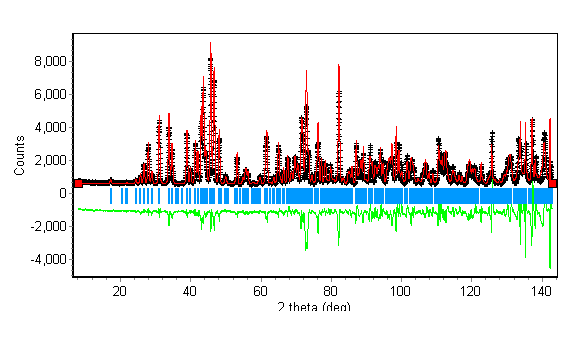
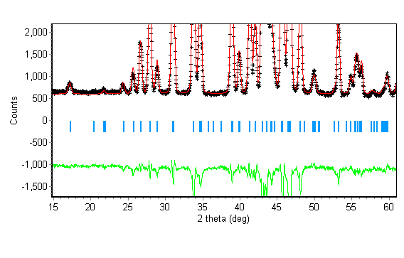
The profile fit at low theta values shows the calculated peaks are at approximately the same theta as the observed peaks, but the intensities are incorrect (Fig. 10). The profile at higher theta shows the calculated peaks are not aligned with the observed peaks (Fig. 11). This is possibly due to incorrect unit cell parameters or unrefined 2q zero error and peak parameters as data set A was collected on a different diffractometer to data set B.
| Fig. 10 |
|---|
| Fig. 11 |
|---|
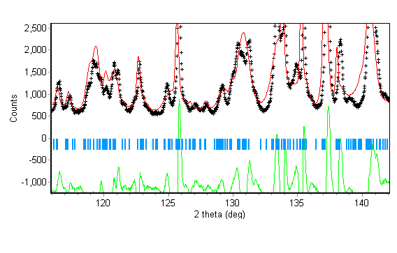
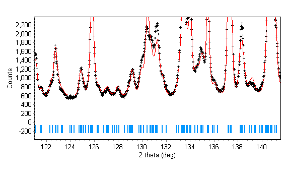
The 2q zero error for the diffractometer at D2B at the ILL, Grenoble, was refined from the starting value of 0.179(13) to 0.2024(13). The resulting R factors were Rp = 13.12, Rwp = 19.60 and Rexp = 2.83. This gave a slightly better fit between calculated and observed peak positions, but fig. 12 shows that the unit cell may need refining to improve the fit.
| Fig. 12 |
|---|
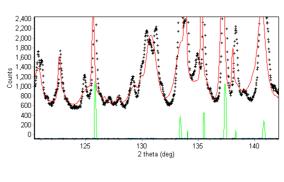
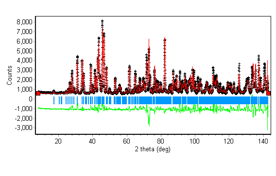
The unit cell was then refined to a = 6.93107(10), b = 8.42162(8) and c = 5.3727(2) and b = 90.153(5)° (Fig. 13). The improvement in peak fit is illustrated by the peaks at 2q values of approximately 125.7 and 137.2° in Fig 13. The R factors refined to Rp = 10.53, Rwp = 14.76 and Rexp = 2.83
| Fig. 13 |
|---|
| Fig. 14 |
|---|
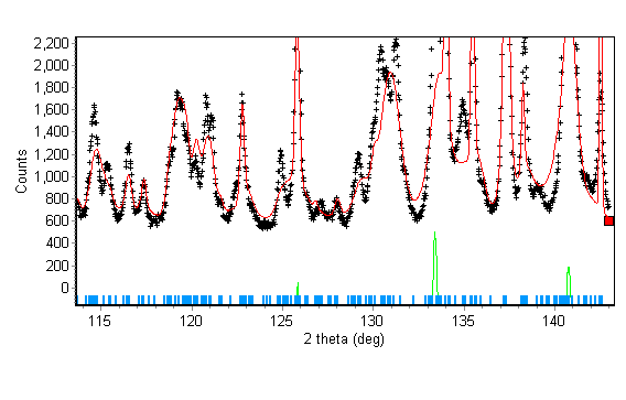
The intensities do not agree between the calculated and observed peaks however. The atomic coordinates were refined successively (Pb followed by the O atoms, and lastly the S atom) , which gave R factors of Rp = 9.32, Rwp = 13.03 and Rexp = 2.82. There were still some discrepancies between calculated and observed peak intensities however.
| Fig. 15 |
|---|
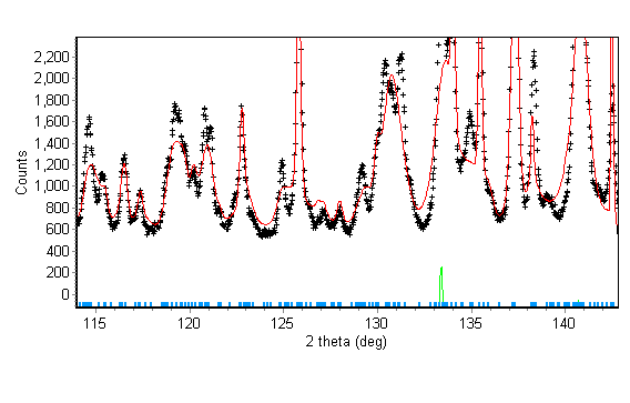
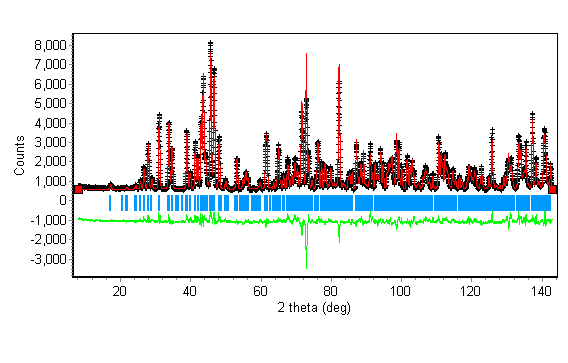
The data in set B was collected on a different diffractometer and so all peak shape parameters were refined. The instrumental parameters U, V and W were refined first, followed by the asymmetry and anisotropic parameters. The R factors refined to Rp = 6.27, Rwp = 8.47 and Rexp = 2.82. The results are shown in Fig. 16 and Fig. 17.
| Fig. 16 |
|---|
| Fig. 17 |
|---|
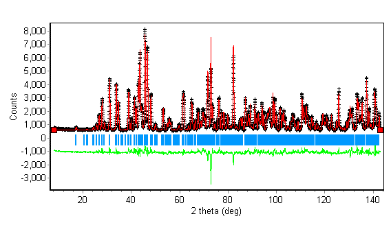
The overall and individual isotropic thermal parameters were then refined to try to improve the calculated intensities. The R factors refined to Rp = 6.01, Rwp = 8.12 and Rexp = 2.28 (Fig. 18 and Fig. 19).
| Fig. 18 |
|---|
| Fig. 19 |
|---|
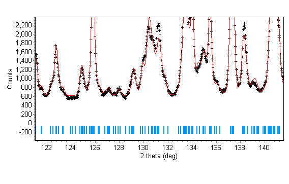
Fig. 20 shows the fit for the two poorest peaks at 2q of about 72.8 and 82.2.
| Fig. 20 |
|---|
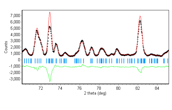
To try to improve the fit the extinction parameter was refined from -0.0031(7) to -0.00063(5). The improvement is shown in Fig. 21. The R factors improved to Rp = 4.22, Rwp = 5.35 and Rexp = 2.82.
| Fig. 21 |
|---|
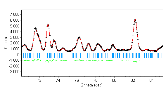
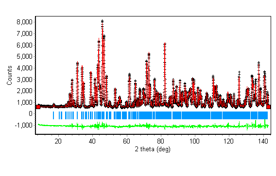
The atoms were then refined anisotropically, giving the profile in Fig. 22 and R factors of Rp = 4.05, Rwp = 5.09 and Rexp = 2.81 where N-P = 5075. c2 = 3.3, where c2=(Rwp/Rexp)2. The following were the final results.
The overall scale factor refined to 2.687(8)
The overall temperature factor refined to 4(5)
The 2q zero error refined to 0.118(2)
The instrumental resolution function parameters refined to U = -0.0441(10), V = -0.153(3), W = 0.205(2)
The peak asymmetry parameter refined to 0.059(3)
The anisotropic peak broadening parameter refined to 0.0273(11)
The extinction parameter refined to -0.00063(5)
The unit cell refined to a = 6.92942(9), b = 8.42053(10), c = 5.37236(8), b = 90.90.2341(7) and V = 313.472(7)
The sample was not corrected for absorption or preferred orientation.
the final profile is shown in Fig. 22 and the fit of the peaks is shown in Fig. 23.
| Fig. 22 |
|---|
| Fig. 23 |
|---|
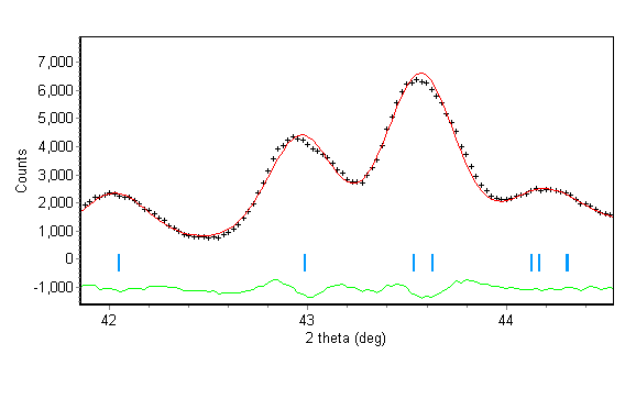
Table 8 shows the final atomic parameters.
Table 8.
| Atom | x | y | z | B11 | B22 | B33 | B12 | B13 | B23 | Biso |
|---|---|---|---|---|---|---|---|---|---|---|
| Pb | 0.16788(13) | 0.18932(8) | 0.2582(2) | 0.00(2) | 0.001(11) | 0.00(3) | 0.00018(12) | 0.0018(2) | 0.0007(3) | -4(4) |
| S | 0.1835(4) | 0.4342(2) | 0.7529(8) | 0.0(2) | 0.00(13) | 0.0(3) | 0.0008(4) | 0.0004(8) | 0.0023(10) | -4(2) |
| O(1) | 0.0930(2) | 0.5922(2) | 0.7610(4) | 0.00(11) | 0.00(8) | -0.0(2) | 0.0005(2) | -0.0013(5) | -0.0007(4) | -3(2) |
| O(2) | 0.0415(2) | 0.3062(2) | 0.7448(4) | 0.00(6) | 0.00(4) | 0.00(10) | -0.0001(2) | -0.0009(2) | -0.0018(4) | -3(4) |
| O(3) | 0.3084(2) | 0.4163(3) | 0.9733(3) | 0.00(10) | 0.00(7) | 0.0(2) | -0.0003(2) | -0.002(2) | -0.0004(3) | -4.1(14) |
| O(3) | 0.3064(2) | 0.4206(3) | 0.5270(3) | 0.0(2) | 0.00(11) | 0.0(3) | -0.0001(2) | -0.002(2) | -0.0016(3) | -4(8) |
Table 9. shows the final molecular dimensions
Table 9.
| Bond lengths in Å | |
|---|---|
| S - O(1) | 1.472(3) |
| S - O(2) | 1.461(3) |
| S - O(3) | 1.4671(4) |
| S - O(4) | 1.490(4) |
| Bond angles in degrees | |
| O(1)-S-O(2) | 112.5(2) |
| O(1)-S-O(3) | 108.5(2) |
| O(1)-S-O(4) | 109.8(3) |
| O(2)-S-O(3) | 110.0(2) |
| O(2)-S-O(4) | 107.8(2) |
| O(3)-S-O(4) | 108.1(2) |
EXPERIMENTAL -DATA SET B ; space group P21/a
The second space group is P21/b with the a axis as the unique axis. Interchanging the a and b axes results in the standard setting of P21/a with the b axis as the unique axis. The second refinement looked at this space group.
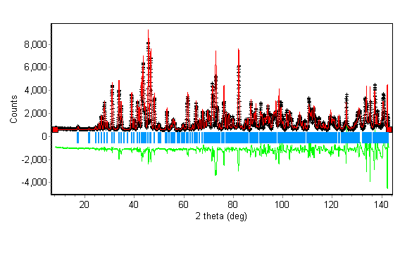
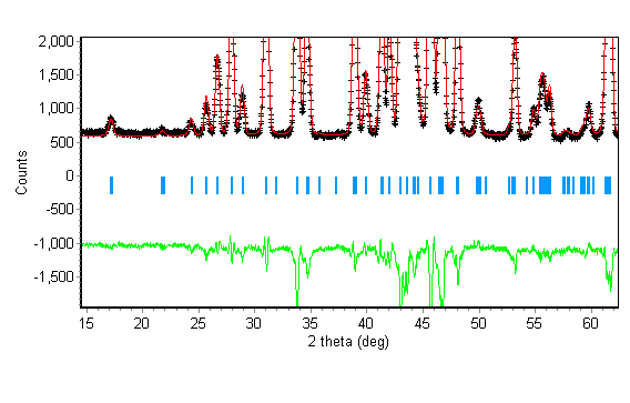
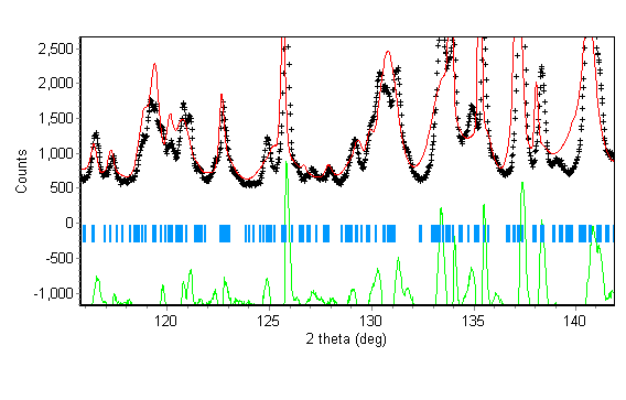
The refinement was carried out as for P21/n. With one cycle of refinement with the scale factor free to refine produced a profile similar to that for P21/n (Fig. 24, Fig. 25 and Fig. 26) with R factors of Rp = 15.86, Rwp = 24.78 and Rexp = 2.83.
| Fig. 24 |
|---|
| Fig. 25 |
|---|
| Fig. 26 |
|---|
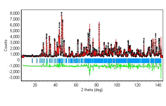
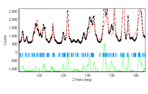
Refining the 2q zero error and the unit cell to try to improve the calculated peak positions resulted in R factors of Rp = 11.18, Rwp = 15.47 and Rexp = 2.83 (Fig. 27 and Fig. 28). The zero error refined to 0.147(2) and the unit cell refined to a = 8.42168(8), b = 6.93104(10), c = 5.3728(2), b = 90.076(8) and V = 313.615(14).
| Fig. 27 |
|---|
| Fig. 28 |
|---|
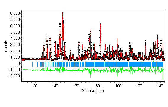
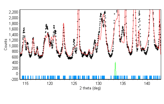
Refining all atomic coordinates resulted in R factors of Rp = 10.26, Rwp = 14.01 and Rexp = 2.82 (Fig. 29 and Fig. 30). Most peak positions seem correct, but there are some which don't fit the model.
The refined coordinates are shown in Table 10.
Table 10.
| Atom | x | y | z |
|---|---|---|---|
| Pb | 0.1889(2) | 0.1658(3)(2) | 0.2476(13) |
| S | 0.4369(9) | 0.1790(10) | 0.782(2) |
| O(1) | 0.5916(4) | 0.0951(5) | 0.767(2) |
| O(2) | 0.3074(5) | 0.0406(5) | 0.7696(14) |
| O(3) | 0.4245(9) | 0.3144(11) | 0.9698(14) |
| O(4) | 0.4154(9) | 0.3011(11) | 0.5287(14) |
| Fig. 29 |
|---|
| Fig. 30 |
|---|
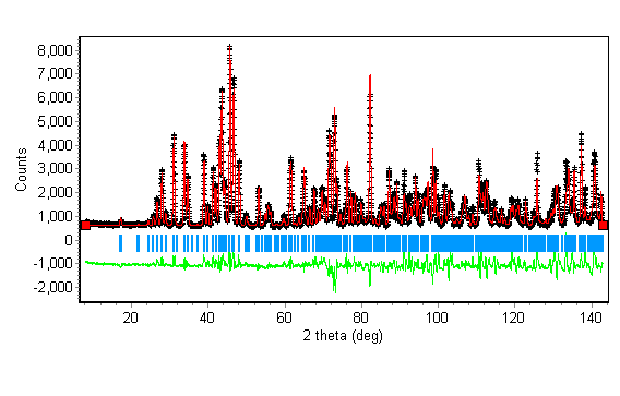
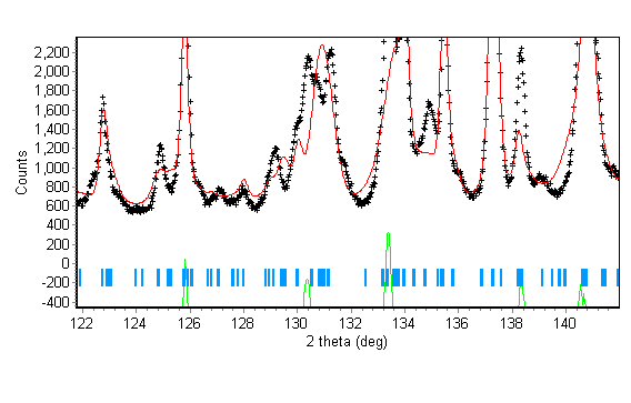
Refining all peak shape and width parameters resulted in R factors of Rp = 9.67, Rwp = 13.24 and Rexp = 2.82 (Fig. 31 and Fig. 32).
| Fig. 31 |
|---|
| Fig. 32 |
|---|
Refining isotropic thermal parameters did not improve the fit , with R factors of Rp = 10.19, Rwp = 16.08 and Rexp = 2.82 (Fig. 33 and Fig. 34). The high angle data still do not fit the calculated profile.
| Fig. 33 |
|---|
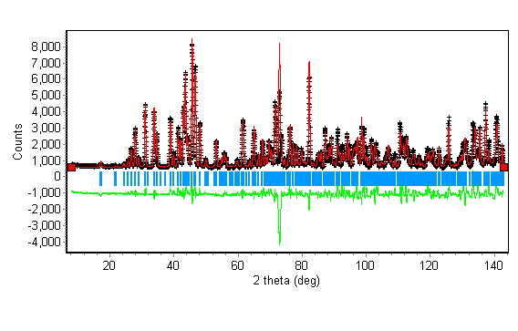
| Fig. 34 |
|---|
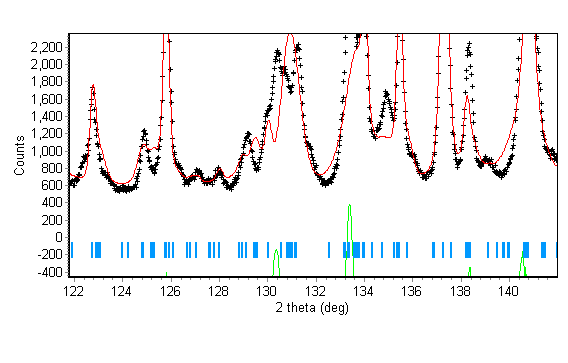
Further refinements of extincton parameters and anisotropic thermal parameters did not produce a better fit between calculated and observed profile, indicating an error in the model (Fig. 35). Thermal parameters did not in fact refine well and the esd's were very large and were not printed out.
The final atomic parameters are in Table 11.
The refined coordinates are shown in Table 10.
Table 11.
| Atom | x | y | z |
|---|---|---|---|
| Pb | 0.1892(3) | 0.1667(4)(2) | 0.2485(11) |
| S | 0.4343(10) | 0.1876(12) | 0.731(4) |
| O(1) | 0.5897(5) | 0.0939(5) | 0.7574(14) |
| O(2) | 0.3081(6) | 0.0414(6) | 0.746(2) |
| O(3) | 0.4235(8) | 0.3137(10) | 0.9703(15) |
| O(4) | 0.4127(10) | 0.2995(10) | 0.524(2) |
| Fig. 35 |
|---|
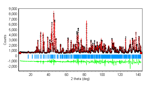
The structure refinement gave the following results, with Rp = 9.50, Rwp = 13.82 and Rexp = 2.81 where N-P = 5079. c2 = 24.2, where c2=(Rwp/Rexp)2.
The overall scale factor refined to 2.686(2)
the overall temperature factor refined to 1(4)
The 2q zero error refined to 0.133(4)
The instrumental resolution function parameters refined to U = -0.0417(3), V = -0.092(9), W = 0.185(5)
The peak asymmetry parameter refined to 0.059(3)
The anisotropic peak broadening parameter refined to 0.0270(9)
The extinction parameter refined to -0.00028(14)
The unit cell refined to a = 8.4210(3), b = 6.9303(2), c = 5.3722(3) and V = 313.52(2)
The sample was not corrected for absorption or preferred orientation.
Table 10. shows the final molecular dimensions for the structure in space group P21/a.
Table 10.
| Bond lengths in Å | |
|---|---|
| S - O(1) | 1.468(9) |
| S - O(2) | 1.471(10) |
| S - O(3) | 1.56(2) |
| S - O(4) | 1.37(2) |
| Bond angles in degrees | |
| O(1)-S-O(2) | 109.5(6)(2) |
| O(1)-S-O(3) | 102.6(12) |
| O(1)-S-O(4) | 116.8(12) |
| O(2)-S-O(3) | 107.4(12) |
| O(2)-S-O(4) | 109.8(13) |
| O(3)-S-O(4) | 110.2(7) |
CONCLUSIONS
On lowering the temperature of the sample of PbSO4, the orthorhombic arrangement of packing is lost. From refinements of both possible monoclinc space groups it was found that the low temperature space group is P21/n with the crystallographic b axis as the unique axis. In the second monoclinc space group, the bond lengths S-O(3) and S-O(4) are long and short respectively, possibly indicating incorrect symmetry elements (ie. space group). All parameters are more precise at lower temperatures due to the smaller thermal displacement about the atomic centres.
CIF FILES
1
data_'PbSO4 - Data Set A'_chemical_formula_structural 'PbSO4'
_symmetry_cell_setting Orthorhombic _symmetry_space_group_name_H_M 'P b n m (equivalent to no. 62)' loop_ _symmetry_equiv_pos_site_id _symmetry_equiv_pos_as_xyz 1 x y z 2 1/2-x, 1/2+y, z 3 1/2+x, 1/2-y, 1/2+z 4 x, y, 1/2-z 5 -x, -y, -z 6 1/2+x, 1/2-y, -z 7 1/2-x, 1/2+y, 1/2-z 8 -x, -y, 1/2+z _cell_length_a 6.9362(13) _cell_length_b 8.4277(15) _cell_length_c 5.3757(10) _cell_angle_alpha 90.0 _cell_angle_beta 90.0 _cell_angle_gamma 90.0 _cell_volume 314.33(9) _cell_formula_units_Z 4 _diffrn_ambient_temperature 293(2) _diffrn_radiation_wavelength 1.594 _diffrn_radiation_type neutron _pd_meas_2theta_range-min 8.0000 _pd_meas_2theta_range_max 62.4750 _pd_meas_2theta_range_inc 0.0250 _computing_rietveld_refinement 'Reitica 1.6.7a' _computing_molecular_graphics 'Reitica 1.6.7a, CorelDRAW 8, Adobe Photoshop' loop_ _atom_site_label _atom_site_type_symbol _atom_site_fract_x _atom_site_fract_y _atom_site_fract_z _atom_site_U_iso_or_equiv _atom_site_thermal_displace_type _atom_site_occupancy Pb Pb 0.1644(11) 0.1894(3) 1/4 -4.3048 Uani 0.5 S S 0.187(3) 0.435(3) 3/4 9.0663 Uani 0.5 O(1) O 0.0903(10) 0.5913(2) 3/4 0.1404 Uani 0.5 O(2) O 0.0444(14) 0.3055(11) 3/4 5.6368 Uani 0.5 O(3) O 0.3051(7) 0.4176(4) 0.9743(8) 7.7544 Uani 1.0 loop_ _geom_bond_atom_site_label_1 _geom_bond_atom_site_label_2 _geom_bond_distance S O(1) 1.47(2) S O(2) 1.47(2) S O(3) 1.467(12) loop_ _geom_angle_atom_site_label_1 _geom_angle_atom_site_label_2 _geom_angle_atom_site_label_3 _geom_angle _geom_angle_site_symmetry_1 _geom_angle_site_symmetry_3 O(1) S O(2) 111.0(15) 1 1 O(1) S O(3) 110.1(11) 1 1 O(2) S O(3)107.5(11) 1 1 O(3) S O(3) 110.6(12) 1 4
2.
data_'PbSO4 - Data Set B'_chemical_formula_structural 'PbSO4'
_symmetry_cell_setting Monoclinic _symmetry_space_group_name_H_M 'P 21/ n (equivalent to no. 14)' loop_ _symmetry_equiv_pos_site_id _symmetry_equiv_pos_as_xyz 1 x y z 2 1/2-x, 1/2+y, 1/2-z 3 1/2+x, 1/2-y, 1/2+z 4 -x, -y, -z _cell_length_a 6.92942(9) _cell_length_b 8.420537(9) _cell_length_c 5.37236(8) _cell_angle_alpha 90.0 _cell_angle_beta 90.2341(7) _cell_angle_gamma 90.0 _cell_volume 313.472(7) _cell_formula_units_Z 4 _diffrn_ambient_temperature 5(1) _diffrn_radiation_wavelength 1.594 _diffrn_radiation_type neutron _pd_meas_2theta_range-min 8.0000 _pd_meas_2theta_range_max 142.9750 _pd_meas_2theta_range_inc 0.0250 _computing_rietveld_refinement 'Reitica 1.6.7a' _computing_molecular_graphics 'Reitica 1.6.7a, CorelPhoto-Paint 8' loop_ _atom_site_label _atom_site_type_symbol _atom_site_fract_x _atom_site_fract_y _atom_site_fract_z _atom_site_U_iso_or_equiv _atom_site_thermal_displace_type _atom_site_occupancy Pb Pb 0.16788(13) 0.18932(8) 0.2582(2) -3.7016 Uani 0.5 S S 0.1835(4) 0.4342(3) 0.7529(8) -3.9351 Uani 0.5 O(1) O 0.0930(2) 0.5922(2) 0.7610(4) -2.9019 Uani 0.5 O(2) O 0.0415(2) 0.3060(2) 0.7448(4) -3.0737 Uani 0.5 O(3) O 0.3084(2) 0.4163(3) 0.9733(3) -4.0877 Uani 1.0 O(4) O 0.3064(2) 0.4206(3) 0.5270(3) -3.6245 Uani 1.0 loop_ _geom_bond_atom_site_label_1 _geom_bond_atom_site_label_2 _geom_bond_distance S O(1) 1.472(3) S O(2) 1.461(3) S O(3) 1.471(4) S O(3) 1.490(4) loop_ _geom_angle_atom_site_label_1 _geom_angle_atom_site_label_2 _geom_angle_atom_site_label_3 _geom_angle _geom_angle_site_symmetry_1 _geom_angle_site_symmetry_3 O(1) S O(2) 112.5(2) 1 1 O(1) S O(3) 108.5(2) 1 1 O(1) S O(4) 109.8(2) 1 1 O(2) S O(3) 110.0(2) 1 1 O(2) S O(4) 107.8(2) 1 1 O(3) S O(4) 108.1(2) 1 1